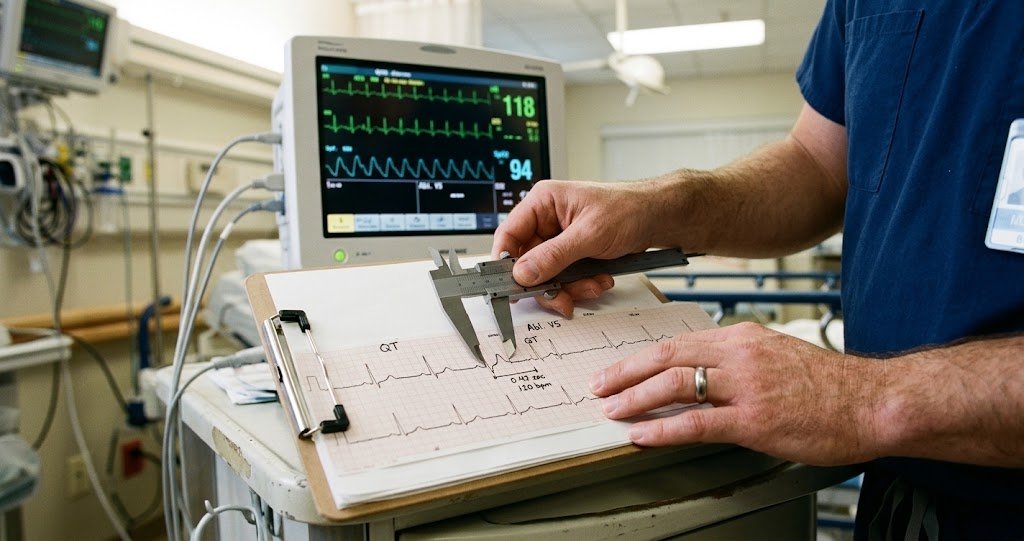
A code doesn’t wait for a provider to work through an ECG strip from scratch. The 8 rhythms every ACLS provider needs to recognize matter less as a memorized list and more as the output of a process — and that process has to run the same way every time, fast enough to finish before the next rhythm check.
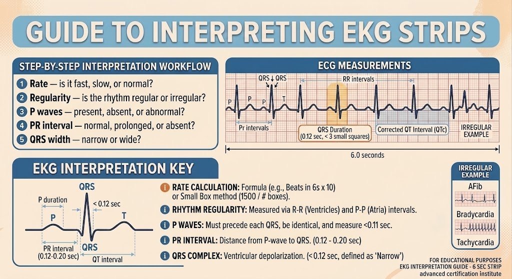
That process is five checks, always in the same order: rate, regularity, P waves, PR interval, QRS width. Run all five and the strip in front of you sorts itself into one of a small number of buckets. Skip the order and default to pattern-matching — “that looks like A-fib” — and the rhythms that are built to be mistaken for each other start winning.
The ACI Method
1. Rate — is it fast, slow, or normal?
Count the number of large boxes between two R waves and divide 300 by that number, or count QRS complexes in a 6-second strip and multiply by 10. Either method gets you close enough in the time it takes to do it. A rate under 60 is bradycardic, 60–100 is normal, over 100 is tachycardic — and that single number already rules out most of the list.
2. Regularity — is the rhythm regular or irregular?
Look at the spacing between R waves across the whole strip, not just two beats. Regular means the spacing stays constant. Irregular splits further: regularly irregular (a repeating pattern, like a fixed conduction ratio) or irregularly irregular (no pattern at all — the hallmark of atrial fibrillation). This one check does more to narrow the list than any other step.
3. P waves — present, absent, or abnormal?
Is there a P wave before every QRS complex? Does it look normal — upright, rounded, consistent shape? A P wave that’s missing, buried, inverted, or shaped differently from beat to beat tells you the impulse isn’t originating from the sinus node the way it should.
4. PR interval — normal, prolonged, or absent?
Measure from the start of the P wave to the start of the QRS complex. Normal is 0.12–0.20 seconds — three to five small boxes. Longer than that signals a conduction delay between the atria and ventricles. No measurable PR interval at all — because there’s no P wave to measure from — rules out sinus-originating rhythms entirely.
5. QRS width — narrow or wide?
Under 0.12 seconds (three small boxes) is narrow, meaning the impulse traveled through the normal conduction pathway. Over 0.12 seconds is wide, meaning it didn’t — either the impulse started in the ventricles themselves, or it took an abnormal path to get there. This is usually the fastest of the five checks and the one that separates the rhythms that can wait from the ones that can’t.
What the Five Checks Rule In and Out
Running the method doesn’t require memorizing which combination equals which rhythm — the five answers narrow the field enough that the identification is usually obvious once you’ve made it that far. Fast and irregularly irregular with no clean P waves points to atrial fibrillation. Fast and regular with a sawtooth baseline instead of P waves points to atrial flutter. Narrow QRS and a rate that lands right around 150 with no visible P waves points to SVT. Wide QRS at a fast rate is ventricular tachycardia until proven otherwise. No organized QRS at all is ventricular fibrillation. A flat line — confirmed in more than one lead — is asystole. An organized rhythm with no pulse is PEA, which the strip alone can’t show you; that one requires checking the patient, not just the monitor.
This is the same territory the last post covered rhythm by rhythm. The difference is the order of operations: that post is the reference for what each rhythm looks like once you know what it is. This is the process for getting there.
Why the Order Matters
Running the checks out of order, or skipping straight to “does this look familiar,” is how sinus tachycardia gets misread as SVT, or atrial flutter gets called atrial fibrillation because both are fast and both look irregular at a glance. The five checks exist specifically to catch the rhythms that resemble each other — that’s the job they’re doing, not just a formality before the obvious answer.
Running them in order also means the strip has already told you the rate, whether it’s regular, and whether the QRS is narrow or wide before you’ve made a single guess. Guessing only enters once — at the end, and by then there’s usually only one rhythm left it can be.
Bottom Line
Five checks, same order, every time: rate, regularity, P waves, PR interval, QRS width. It takes about a minute the first few times through and considerably less once it’s automatic — which is the point. The rhythm on the monitor determines what happens next in a code, and the ACI Method is how a provider gets from strip to rhythm to action without guessing in between.
Advanced Certification Institute (ACI) provides online ACLS, PALS, and BLS certification and recertification for healthcare professionals, built around the current AHA/ILCOR guidelines. Explore ACI’s ACLS courses.