A code team doesn’t have time to think through an ECG strip from first principles. The rhythm has to be recognized in seconds, because the next action — compressions, a shock, a drug, or nothing at all — depends entirely on getting it right. ACLS certification tests this recognition directly, but the real reason it matters isn’t the exam. It’s that the ACLS Cardiac Arrest Algorithm branches into two completely different treatment paths based on which rhythm is on the monitor, and picking the wrong branch costs a patient the seconds that actually save a life.
The eight rhythms below are the ones every ACLS provider needs to identify on sight, not work out. Four of them are the arrest rhythms the algorithm is built around. The other four show up constantly outside of arrest and matter because they’re commonly mistaken for something more dangerous than they are — or the reverse.
1. Normal Sinus Rhythm
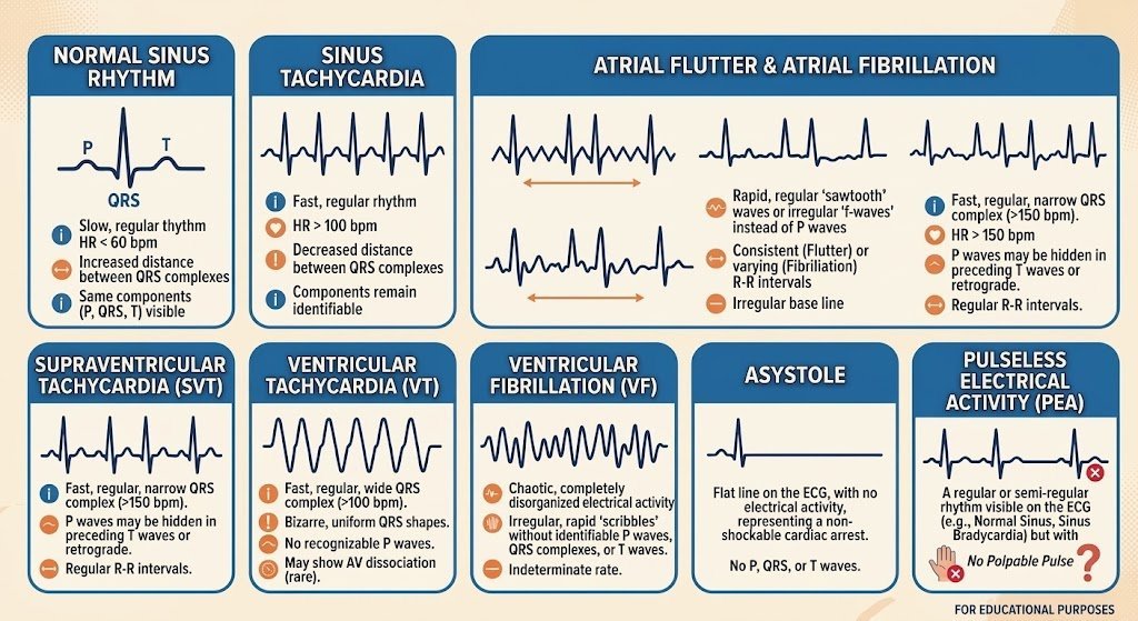
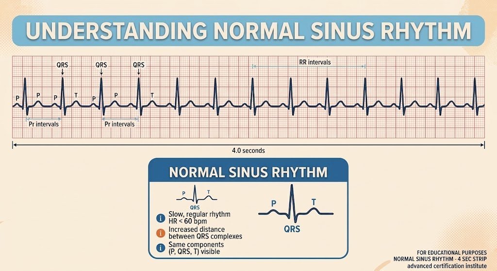
What it looks like: Rate 60–100 bpm, regular, a P wave before every QRS complex, PR interval 0.12–0.20 seconds, QRS complex under 0.12 seconds.
Why it matters: This is the reference point. Every other rhythm on this list is defined by how it deviates from sinus rhythm — a faster rate, an irregular rhythm, a missing P wave, a widened QRS. Providers who can’t identify normal sinus rhythm confidently will struggle to identify what’s abnormal about anything else.
What to do: Nothing. It’s the target rhythm, not a finding that requires intervention.
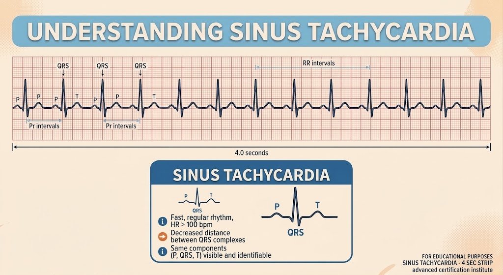
2. Sinus Tachycardia
What it looks like: Rate above 100 bpm, regular, normal P waves before each QRS, narrow QRS complex.
Why it matters: Sinus tachycardia is a response, not a primary arrhythmia. Fever, pain, hypovolemia, hypoxia, anxiety, and pulmonary embolism all produce it. Treating the rate instead of the cause is the most common mistake made with this rhythm — a patient tachycardic from blood loss doesn’t need a rate-control drug, they need volume.
What to do: Identify and treat the underlying cause. The rhythm itself isn’t the problem.
3. Atrial Fibrillation and Atrial Flutter
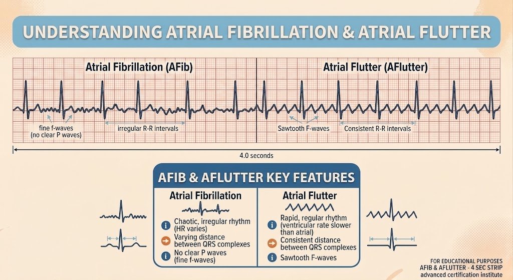
What it looks like: Atrial fibrillation is irregularly irregular, with no discernible P waves and a chaotic fibrillatory baseline; QRS is usually narrow. Atrial flutter looks different and shouldn’t be mistaken for it: a distinctive sawtooth pattern of atrial activity, often regular, typically conducting to the ventricles at a fixed ratio — 2:1 is common, producing a ventricular rate that lands suspiciously close to 150 bpm.
Why it matters: These are the two most common sustained atrial arrhythmias providers will encounter, and they get confused for each other more than the strip difference should allow. Both carry a real stroke risk from blood stasis in the atria, and both can cause hemodynamic instability on their own if the ventricular rate runs fast enough. The sawtooth pattern is the tell that separates flutter from fibrillation at a glance — worth training the eye to catch it specifically, not just default to “irregular rhythm, must be A-fib.”
What to do: Rate or rhythm control for either, based on the patient’s stability and how long the arrhythmia has been present. An unstable patient with new-onset A-fib or flutter gets synchronized cardioversion — flutter in particular tends to respond well to cardioversion at lower energy than fibrillation typically requires.
4. Supraventricular Tachycardia (SVT)
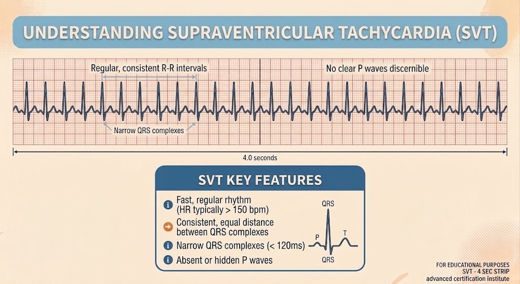
What it looks like: Rate typically 150–250 bpm, regular, narrow QRS, P waves usually absent or buried in the preceding T wave.
Why it matters: SVT gets confused with sinus tachycardia at a glance and with ventricular tachycardia when the rate is high — and the treatment for each is different enough that misreading the strip leads to the wrong intervention. SVT originates above the ventricles, which is exactly why vagal maneuvers and adenosine work on it and don’t touch a ventricular rhythm.
What to do: Vagal maneuvers first if the patient is stable, then adenosine. An unstable patient goes straight to synchronized cardioversion.
5. Ventricular Tachycardia (VT)
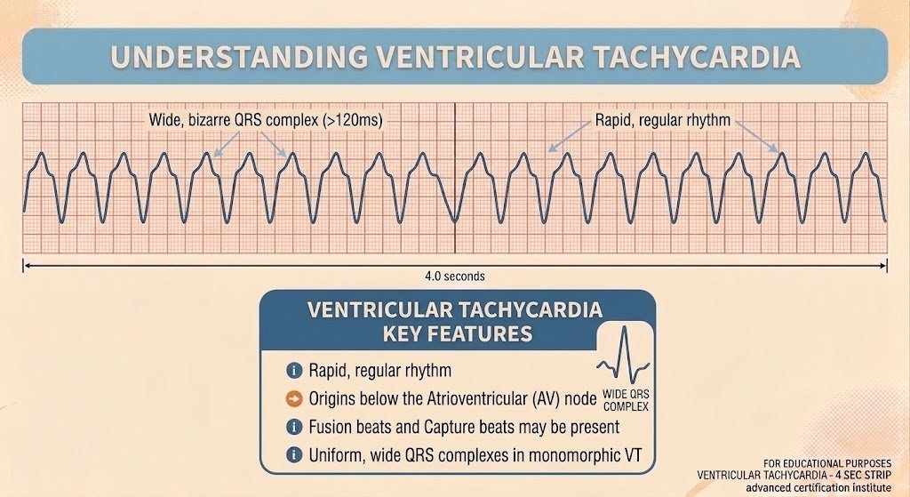
What it looks like: Wide QRS complex (over 0.12 seconds), rate typically 150–250 bpm, can be regular or irregular, P waves absent or dissociated from the QRS.
Why it matters: VT is the rhythm where the pulse check decides everything. Pulseless VT is treated exactly like ventricular fibrillation — it’s a shockable arrest rhythm. VT with a pulse is a different problem entirely: an unstable patient needs synchronized cardioversion, a stable one gets an antiarrhythmic. Same waveform, three different treatment paths, depending on one thing the strip alone can’t tell you.
What to do: Check for a pulse before anything else. Pulseless VT: defibrillate immediately, resume CPR, follow the arrest algorithm. VT with a pulse, unstable: synchronized cardioversion. VT with a pulse, stable: amiodarone or procainamide.
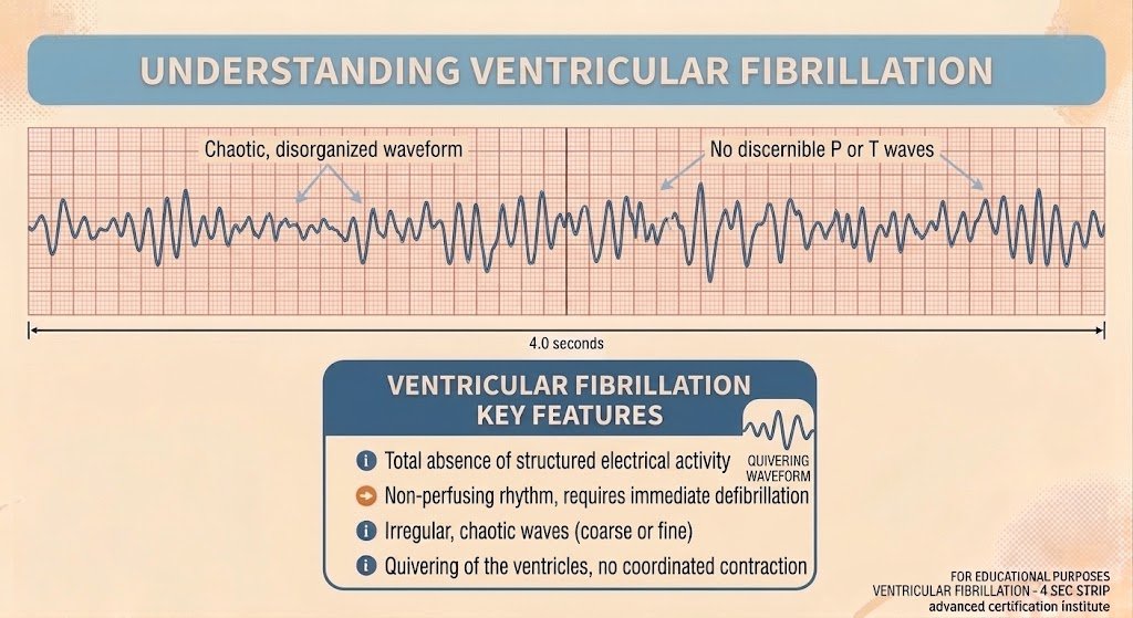
6. Ventricular Fibrillation (VF)
What it looks like: Chaotic, disorganized waveform with no identifiable QRS complexes at all.
Why it matters: VF never produces a pulse. There’s no ambiguity here the way there is with VT — if it’s VF, the patient is in cardiac arrest, and every second before defibrillation lowers the odds of a successful outcome.
What to do: Immediate defibrillation. CPR before and after the shock, epinephrine, and consider amiodarone or lidocaine per the algorithm if VF persists after the first shock.
7. Asystole
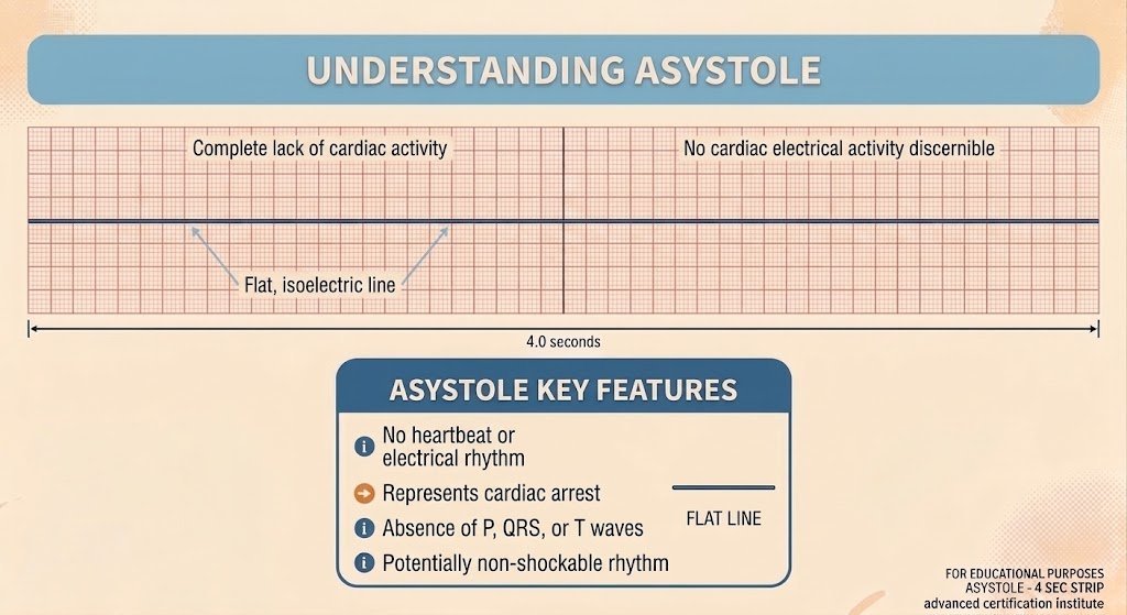
What it looks like: A flat line. No electrical activity at all.
Why it matters: This is the rhythm most likely to trigger the wrong reflex. Asystole is not shockable, and shocking it does nothing — there’s no organized electrical activity for a defibrillator to interrupt. Before calling it, confirm the flatline in more than one lead and check that the leads are actually connected; a disconnected lead looks identical to asystole on the monitor.
What to do: CPR and epinephrine, not defibrillation. Work through the H’s and T’s — the reversible causes of arrest — because asystole rarely resolves without finding and correcting one of them.
8. Pulseless Electrical Activity (PEA)
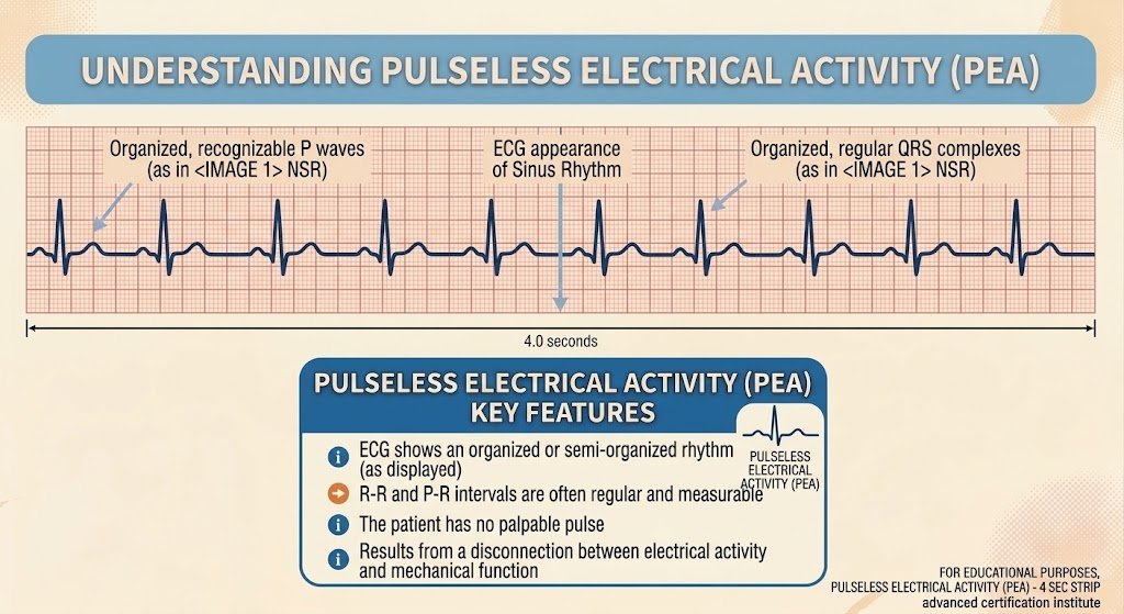
What it looks like: An organized rhythm on the monitor — sometimes one that looks entirely normal — with no palpable pulse.
Why it matters: PEA is the rhythm that looks like it should be working and isn’t. The electrical system is firing correctly; something else — hypovolemia, hypoxia, tension pneumothorax, cardiac tamponade, a massive pulmonary embolism, or another reversible cause — is preventing that electrical activity from producing a mechanical contraction that generates a pulse.
What to do: CPR and epinephrine, same as asystole — it’s non-shockable. The real work is identifying the reversible cause fast enough to correct it before the arrest becomes unrecoverable.
Where This Fits in the Algorithm
Rhythms 5 through 8 are the ones the ACLS Cardiac Arrest Algorithm is built around, and they split into exactly two treatment paths. VF and pulseless VT are shockable — the algorithm calls for defibrillation. Asystole and PEA are not — the algorithm calls for CPR, epinephrine, and a search for a reversible cause instead. Everything else in the algorithm sits downstream of that first branch point, which is why recognizing which side of it a rhythm falls on has to be automatic, not something worked out mid-code.
Bottom Line
Eight rhythms account for nearly everything an ACLS provider needs to recognize without hesitation: the sinus baseline, three rhythms that mimic danger more than they cause it, and four arrest rhythms that split into a shockable path and a non-shockable one. Knowing the name of a rhythm matters less than knowing which action it triggers — that’s the actual skill the algorithm is testing.
Advanced Certification Institute provides 100% online BLS certification and recertification for healthcare professionals — built for healthcare workers, by healthcare workers. Explore our courses.
Use code BLOGREADER for a discount off your next order!
Advanced Certification Institute (ACI) provides online ACLS, PALS, and BLS certification and recertification for healthcare professionals, built around the current AHA/ILCOR guidelines. Explore ACI’s ACLS courses.